General Protocols for Material Preparation, PCR and Restriction Digestion
PCR-RFLP Identification of Globodera pallida, G. rostochiensis, Heterodera spp. and Meloidogyne spp.
This manual provides technical information on the used of PCR-RFLP for Globodera pallida, G. rostochiensis, Heterodera spp., and Meloidogyne spp. identification. The techniques presented here are intended as a general guide, and are the ones best suited to our own needs at the Nematology Laboratory, Plant Pest Diagnostics Branch, California Department of Food and Agriculture.
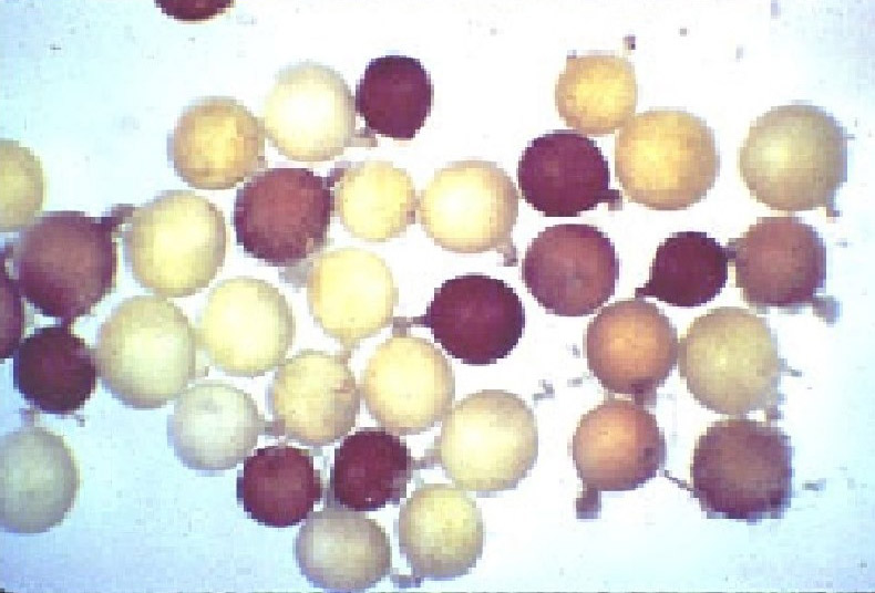
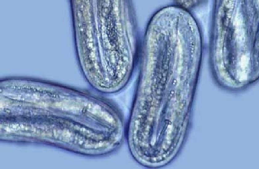
In the CDFA Nematology Laboratory, plant parasitic nematodes are extracted from soil samples using combined techniques of 60 mesh gravity sieve, sugar centrifugation, and misting method. Nematodes are examined using dissection microscopes that allow preliminary assignment to genus. Globodera, Heterodera and Meloidogyne J2s are further identified by light microscopy on temporary glass slides. The preliminary identified nematode eggs, infective juveniles, males and/or young females of Globodera, Heterodera and Meloidogyne are analyzed further with molecular techniques.
Make records in the lab note to include the laboratory test number, pest and damage record (PDR) number, host, location/county, and sampling/testing date. Label the 0.5 ml PCR tube with the testing number, and match the number with the PDR number in the lab note.
The pre-identified nematode eggs, juveniles, males and/or young females are placed in a drop of 40ml 0.1M Tris-HCl (pH8.0) on a glass slide. The nematodes are smashed with a dental file to release the nematode body contents, and then transferred directly to the bottom of a 0.5ml PCR reaction tube. A 10 ml portion of the nematode solution serves as DNA template for PCR reaction. Store the tubes at -20°C. They should remain usable for several weeks.
For a single 25μl PCR reaction, the components are listed in Table 1.
Table1: PCR component for nematode species identification.
| Template DNA | 10μl | Smashed nematode samples in the PCR tube |
|---|---|---|
| Tris pH8.0 | 2.5μl | |
| 10x Buffer | 2.5μl | Promega, Inc. |
| MgCl2 | 3μl | Promega, Inc. |
| dNTP | 0.5μl | 200µM of each dNTP final concentration |
| Primer 1&2 mix | 4μl | 0.36µM of each primer final concentration |
| BSA | 2μl | |
| Taq | 0.5μl | ~2.0 units final amount |
The master mix is made according to the number of samples tested include enough for a positive and negative control. A negative control is a 15.0μl of master mix with 10.0μl of 0.1M Tris-HCl buffer. A positive control is a 15.0μl of master mix with 10.0μl of a control DNA template that is known to amplify consistently.
The thermo-cycles are programmed in general temperature parameters as follow:
After step #3, the temperature on the thermo-cycler is programmed at 4˚C forever.
1.2% agarose gel in 1X TAE buffer is used to separate the PCR products. 5.0μl PCR product from each sample is loaded into the gel with 100bp and 1kb DNA ladders. The agarose gel is run at constant voltage (80V) and stained in EtBr for 10 min. Rinse the gel off several times with H2O, and look at it on the UV box. Take a picture of the gel and label the picture with the test number and date. Tape the gel picture in the lab note as a record.
Restriction digestions are usually required for nematode species identifications. About 5μl PCR product will provide enough DNA, when it is digested, to show each small fragment well on the agarose gel. Digestions seem to work the best when the total volume is about 10-15μl. This total includes DNA volume, 10X buffer, enzyme, and maybe some ddH2O (Table 2).
Table 2: A typical restriction component for nematode identifications.
| A typical restriction component for nematode identifications. | |
|---|---|
| 5μl | PCR product |
| 1μl | 10x enzyme buffer* |
| 1μl | restriction enzyme |
| 3μl | H2O |
| 10μl | total volume |
Place the tubes into a micro-rack, and place the rack in the proper incubation conditions, usually at 37ºC in an incubation oven. The tubes are allowed to incubate for one to two hours.
A good quality agarose like MetaPhor or Synergel works best for small size DNA fragments. To separate the digested DNA fragments, which can be small (100bp-300bp) in size, the agarose gel concentration can be increased to 2%.
Nematode Species Identification
PCR-RFLP Identifications for Globodera pallida, and G. rostochiensis

There are two PCR testing protocols for Globodera pallida and G. rostochiensis currently used at CDFA nematology laboratory. The nematode species are identified based on the PCR or PCR-RFLP fragment sizes.
The first PCR test is conducted with primers rDNA2 (5’ TTGATTACGTCCCTGCCCTTT 3’) and rDNA1.58S (5’ACGAGCCGAGTGATCCACCG 3’) located in the ITS-1 regions of cyst nematodes, 18s and 5.8s genes respectively (Fleming, et. al., 1998, Aspects of applied biology, protection and production of sugar beet and potatoes. 52:375-382). After the PCR products are separated on a gel, the Globodera spp. can be pre-identified, both nematode species, G. rostochiensis and G pallida, produce the PCR bands of 755bp. (The common Heterodera species usually produce an 800bp band). The restriction enzyme Hinf 1 will be used to differentiate G. rostochiensis and G pallida. The digestion products for G. rostochiensis are 233bp + 522bp. If the nematode is G pallida, Hinf 1 restriction bands will be 152bp, 233bp, and 370bp.
If a certain nematode isolate is tested with first PCR protocol and found to be G. pallida or G. rostochiensis. The second PCR protocol will be used to confirm the identification. The second PCR test is conducted also in ITS1 region with species specific primer set ITS5 (5’ GGAAGTAAAAGTCGTAACAAGG) and PITSp4 (5’ACAACAGCAATCGTCGAG 3’) for G. pallida; and primer set ITS5 and PITSr3 (5’ AGCGCAGACATGCCGCAA 3’) for G. rostochiensis (Bulman and Marshall, 1997. New Zealand Journal of Crop and Horticultural Science. 25:123-129). The PCR reaction from ITS5/PITSr3 will positively confirm G. rostochiensis and the PCR product will be 435bp. The PCR reaction from ITS5/PITSp4 will positively detect G. pallida and the PCR product will be 266bp.
No golden cyst nematode G. rostochiensis and/or potato cyst nematode G. pallida have been found in California.
PCR-RFLP Identifications for Heterodera spp.

Due to the multiple cyst-forming nematode species in the genus Heterodera, both the ITS1 and ITS2 regions are chosen for PCR reactions to include the maximum information for species identifications. The PCR products from Primers AB 28 (5'ATATGCTTAAGTTCAGCGGGT3') and TW 81 (5'GTTTCCGTAGGTGAACCTGC3') will amplify a PCR product approximately 1060bp that include both ITS1 and ITS2 from the common species of Heterodera. The PCR products are then digested with different restriction enzymes for species differentiations. (Subbotin, S. A. et al. 2000. Nematology. 2(2) 153-164).
The most common cyst forming nematode in California is sugar beet cyst nematode Heterodera schachtii. The restriction enzyme MvaI is used and the digestion products include a few DNA fragments, e. g., 1010bp, 840bp, 760bp, 630bp, 220bp, 150bp, and 80bp. H. cruciferae can be differentiated from H. schachtii by the same MvaI cut with the products of 760bp, and 300bp. For detailed and complete information about other common species of Heterodera, please review Dr. Subbotin's publication (Nematology. 2(2) 153-164.)
PCR-RFLP Identifications for Meloidogyne spp.Globodera sp.

The PCR amplification for Meloidogyne spp. is conducted with two primers located in the COII and 16S ribosomal mitochondrial genes respectively (Powers and Harris, JON 25(1): 1-6; Stanton, et al., Fundam. Appl. Nematol. 20(3): 261-268). According to the available published information, at least eight root-knot nematode species (Meloidogyne spp.) can be identified with PCR-RFLP fragment sizes from this mitochondrial DNA, namely: M arenaria, M. chitwoodi, M. graminis, M. hapla, M. incognita, M. javanica, M. mayaguensis, and M. partityla. The C2F3 primer (5’ GGTCAATGTTCAGAAATTTGTGG 3’) from Powers is chosen as up-stream primer, and the MRH106 from Stanton's publication is used as down-stream primer (5’ AATTTCTAAAGACTTTTCTTAGT 3’) in the CDFA nematology laboratory for this purpose.
Based on the size of amplification PCR (or PCR-RFLP) products, some of the most common Meloidogyne spp can be identified as follow:
- The amplification product of ~1,300bp is designated as M. arenaria.
- The amplification products of approximately 1,800bp are further digested with HinfI.
- If products of 1400bp and 400bp are produced the specimen is designated M. incognita.
- If no digestion occurred the specimen is designated M. javanica.
- If the amplification products are about 600bp. The PCR products are subjected to a DraI digestion:
- If the digestion products are 258bp, 119bp, 40bp, 18bp, and 156bp, the species is M. chitwoodi.
- If the digestion products are 246bp, 198bp, 51bp and 103bp; or 444bp, 51bp and 103bp due to a single nucleotide mutant, the species is M. hapla.
In addition, M. marylandi, M. nataliei, and M. nassi were reported to produce the ~600bp DNA bands, but no DraI cutting sizes were available from publications.
Due to the common occurrence of M. chitwoodi and M. hapla in California and both species are important in regulatory nematology, a second PCR test of the IGS region is conducted to confirm the species identification (Wishart, et al. Phytopathology 92:884-892). In this PCR test, a universal primer JMV1 (5' GGATGGCGTGCTTTCAAC 3') and two species specific primers are used for M. chitwoodi JMV2 (5' TTTCCCCTTATGATGTTTACCC 3'), and M. hapla JMVhapla (5' AAAAATCCCCTCGAAAAATCCACC 3'). The reaction can be conducted in a multiplex PCR, the product of M. chitwoodi is 540bp and M. hapla to be 440bp. In addition to the PCR tests, when root galls are available in some samples, Meloidogyne adult females are isolated for morphological and isozyme analyses as supplementary techniques for identification.
References
- Bulman, S. R., and Marshall, J. M. 1997. Differentiation of Australasian potato cyst nematode (PCN) populations using the polymersare chain reaction (PCR). New Zealand Journal of Crop and Horticultural Science. 25: 123-129.
- Fleming, C. C., Turner S. J., Powers, T. O, and Szalanski, A. L. 1998. Diagnostics of cyst nematodes: use of the polymerase chain reaction to determine species and estimate population levels. Aspects of applied biology, protection and production of sugar beet and potatoes. 52:375-382.
- Powers, T. O., and Harris, T. S. 1993. A polymerase chain reaction method for identification of five major Meloidogyne species. Journal of Nematology 25(1): 1-6.
- Stanton, J., Hugall, H., and Moritz, C. 1997. Nucleotide polymorphism and an improved PCR-based mtDNA diagnostic for parthenogenetic root-knot nematodes (Meloidogyne spp.) Fundamental and Applied Nematology 20(3): 261-268.
- Subbotin, S. A., Waeyenberge, L., and Moens, M. 2000. Identification of cyst forming nematodes of the genus Heterodera (Nematoda: Heteroderidae) based on the ribosomal DNA-RFLP. Nematology. 2:153-164.
- Szalanski, A. L., Sui, D. D., and Powers, T. O., 1998. Globodera pallida 18S ribosomal RNA gene, partial sequence; internal transcribed spacer 1, complete sequence; and 5.8S ribosomal RNA gene, partial sequence. NCBI GenBank. AF016871.
- Szalanski, A. L., Sui, D. D., and Powers, T. O., 1998. Globodera rostochiensis 18S ribosomal RNA gene, partial sequence; internal transcribed spacer 1, complete sequence; and 5.8S ribosomal RNA gene, partial sequence. NCBI GenBank. AF016878.
- Wishart, J., Philips, M.S., and Blok, V. C. 2002. Ribosomal intergenic spacer: a polymerase chain reaction diagnostic for Meloidogyne chitwoodi, M, fallax, and M. hapla. Phytopathology 92: 884-892.
Report a Pest
Nematology Sub-Menu
Plant Pest Diagnostics Center
CDFA Collections
General Info & Links
Quick Links
Contact Us
Plant Pest Diagnostics Center
3294 Meadowview Road
Sacramento, CA 95832
Phone: (916) 262-1100
Fax: (916) 262-1190
Map & Directions
